X-ray radiographic system
X-ray radiography is a method to reveal internal structure of objects. The examined object is illuminated with a beam of X-rays from a generator. X-rays are absorbed by the object depending on its density and composition. The attenuated X-ray beam is then detected in an imaging X-ray camera. In other words, a shadow in the X-ray beam cast by the object is detected.

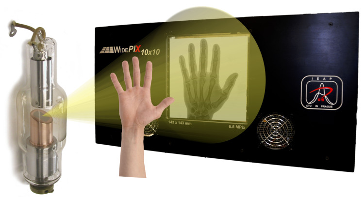
Illustration of the radiography principle: the X-ray source (left) generates beam of X-ray photons. The beam is cast onto the sample and the sample projects a shadow onto the imaging detector. The detector records the image and transfers it into a computer.
The X-ray radiography principle explained above was used already by Wilhelm Conrad Röntgen in 1896. Hence, the principle is more than 100 years old. What has been tremendously improved since the 19th century are both, the X-ray sources and X-ray cameras. This website focuses on the sensor part of the X-ray imaging chain.
The imaging systems presented on this website are single photon counting pixel detectors. These devices represent the cutting edge of current radiation imaging technology. The term “single photon counting” means that every single photon of X-ray radiation detected in individual pixels is processed and counted. The processing of individual photons brings two major advantages:
- low noise in images,
- energy sensitivity.
The photon counting principle of detection eliminates all other sources of noise that are present in CCD or flat-panel based cameras. This leads to considerably better signal-to-noise ratio and therefore detectability of more details in images.
The energy sensitivity is as important advance in the imaging technology as was the colour photography and film. Contrary to regular X-ray imaging cameras, the photon-counting devices can discriminate or even directly measure energy (wavelength) of incoming photons. Since each element has different X-ray attenuating properties, it is possible to estimate even material composition of the sample if the energy of photons is measured. This is a major improvement of X-ray imaging systems thanks to these new cameras.

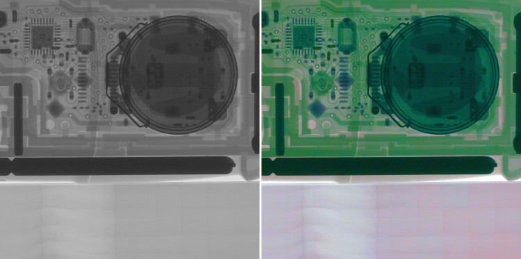
Comparison of a regular X-ray image and “colour” X-ray image where the colours correspond to different X-ray energy channels. Different materials in the electronics of a car key are easily distinguished.


